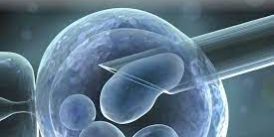
In-vitro fertilization (IVF) and Preimplantation Genetic Testing (PGT) in sickle cell disease and family balancing
Assisted Reproductive Technology development started after the breakthrough of Patrick Steptoe and Robert Edwards in the successful birth of a baby in 1978, after in-vitro fertilization and embryo transfer (IVF-ET). Increase knowledge in the Assisted Reproductive Technology was in the development of Preimplantation genetic diagnosis. We in Nigeria were also at the forefront of catching up with these technologies. We established and reported success with the PGT technique for sickle cell carriers. Recognizing that Sickle cell disease carriers are present in our population, we utilized these techniques to help sickle cell carrier couples have normal sickle cell babies Nigeria Medical Journal 2014). The utilization of the procedure in our environment can eliminate sickle cell disease over a long time. As we all know, SCD is a group of disorders that affects hemoglobin, the molecule in red blood cells that delivers oxygen to cells throughout the body. SCD patients have a typical hemoglobin molecule called hemoglobin S, which distorts the red blood cell’s original biconcave disc to a sickle or crescent shape. This article discusses the roles of IVF procedures in curing SCD.
How does IVF relate to SCD?
In vitro fertilization, popularly known as IVF,https://www.mayoclinic.org/tests-procedures/in-vitro-fertilization/about/pac-20384716 is an assisted reproductive technique that achieves fertilization by retrieving oocytes from the female, collecting sperm from the male, and then combining them manually in a laboratory dish through a series of the procedure. From the first success in 1978, there have been upgrades in IVF procedures and newer techniques like ICSI, PGT, and PCR, among many others. The introduction of these procedures gave rise to exploring knowledge into the viability and quality of embryos both morphologically and genetically; one such is the Pre genetic implantation testing as both a preventive and corrective procedure for SCD.According to the World Health Organization (WHO), sickle cell disease is the most prevalent genetic disease in the African region.https://www.afro.who.int/health-topics/sickle-cell-disease While 75% of patients with SCD live in sub-Saharan Africa, Nigeria alone accounts for more than 100,000 births each year. It cannot but be overemphasized that sickle cell disease is fast becoming an African disease.Nigeria experienced her first IVF success by our very own Prof Oladapo Ashiru and Prof Osato Giwa-Osagie in 1989 at the Lagos University Teaching Hospital,https://www.ncbi.nlm.nih.gov/pmc/articles/PMC4071672/ making it the first in Sub-Saharan Africa. Since then, there has been a rise in the awareness of IVF among Nigerians and an increase in the number of fertility clinics in Nigeria, collectively working to solve the problems of infertility. A follow up to this development is establishing additional technologies in Assisted Reproductive Technology like Preimplantation Genetic Testing (PGT)..
The history of ART dates back to 1969 when Yanagimachi and Chang reported capacitation for in-vitro fertilization of hamster eggs. Capacitation, a term used to describe hyper activated motility of the sperm, is required for fertilization to take place.1 Following this, in 1977, Andrew Schally and Roger Guillemin were awarded the Nobel Prize in Medicine and Physiology for their work in the isolation of LHRH from the hypothalamus.2
Roger Guilleman and Andrew V Schally
In 1978, Ashiru and Blake also reported FSH positive feedback mechanism on the pituitary.3 These and many others have been the bedrock for the achievement of the first live birth via in vitro fertilization (IVF), Louis Brown, popularly called the first ‘test-tube’ baby in 1979.4 This ground breaking success was achieved by Steptoe and Edward in London and Robert Edwards was subsequently awarded the Nobel Prize in Medicine in 2010.
Patrick Steptoe and Robert Edwards 1978.
Other countries reported their successes, Australia by Carl Woods in 1980, 5 USA by Howard & Georgeanna Jones in 1982 6 and Nigeria by Oladapo Ashiru, Osato Giwa-Osagie in 1984 7 and the delivery of a baby through IVF in 1989. 8
FIGURE 3 O. F. Giwa-Osagie, R. Edwards and O. A. Ashiru 1992
WHAT IS IVF?
IVF means fertilization achieved outside of the body.https://www.mayoclinic.org/tests-procedures/in-vitro-fertilization/about/pac-20384716 It involves ovulation induction, oocyte retrieval, sperm preparation, oocyte stripping, insemination and fertilization in a culture dish and finally embryo transfer. Oocyte retrieval and embryo transfer processes are usually done under ultra-sound guidance. Also, it is important to know that gamete handling is usually done under strict temperature control.https://medicalartcenter.com/in-vitro-fertilisation-ivf/ Without the development of IVF, pre-implantation genetic diagnosis (PGD) would not have been possible.
Illustration of Steps involved in IVF
WHAT IS PGD?
PGD as the name implies involves testing for specific genetic defects in the DNA code prior to embryo implantation. Usually, there is targeted testing of a known genetic abnormality in the couple.
Preimplantation genetic diagnosis (PGD) is diagnosis of a genetic condition prior to achievement of a pregnancy. PGD was first performed in the early 1990’s as a way for couples to prevent the pregnancy of a child with a genetic disease. Currently at Medical Art Center, we are able to offer PGD testing for genetic conditions including sickle cell anaemia and chromosome aneuploidies. We work in collaboration with Genesis Genetics, a world renowned genetics institute. They pioneered PGD testing of embryos for inherited genetic abnormalities.
DOES PGD REPLACE PRENATAL TESTING?
No, PGD does not replace prenatal testing such as chorionic villus sampling (CVS) or amniocentesis. PGD is a research based test allowing for a similar diagnosis to those available by prenatal testing. However, prenatal testing is still recommended as these widely used and well accepted prenatal tests remain the gold-standard of modern obstetrics and are necessary in this setting should a pregnancy ensue
HISTORY OF PGD
Edwards and Gardner successfully performed the first known embryo biopsy on rabbit embryos in 1968. 9 In humans, PGD was developed in the United Kingdom in the mid-1980s as an alternative to current prenatal diagnoses. Initially, PGD revolved around determination of gender as an indirect means of avoiding an X-linked disorder.https://ivf-worldwide.com/cogen/oep/pgd-pgs/history-of-pgd-and-pgs.html In 1989 in London, Handyside and colleagues reported the first unaffected child born following PGD performed for an X-linked disorder.10
Kemi Ailoje, Alan Handyside, Oladapo Ashiru and Rose Ogbeche
Yuri Verlinsky et al 1999 established Polar body testing. 11 PGD became increasingly popular during the 1990s when it was used to determine a handful of severe genetic disorders, such as sickle-cell anemia, Tay Sachs disease, Duchenne’s muscular dystrophy, and Beta-thalassemia. In 2010 Oladapo Ashiru et al used PGD with FISH (Fluorescent in situ Hybridization) for chromosomal abnormalities and family balancing and in 2013 published their first success with the use of PGD to deliver a sickle cell free baby boy. 12(NMJ 2014)
As of 2006, more than 15,000 PGD cycles have been reported. PGD is currently available for most known genetic mutations. Although the indications for PGD are well established, it is a relatively new, and evolving technique.
INDICATIONS FOR PGD
Autosomal dominant disorders: This is situation where one of the couple is a carrier of the genetic defect (e.g., Huntington Disease or dwarfism)
Autosomal recessive disorders: This is a situation whereby both couple are carriers of the genetic defect (e.g., Sickle Cell Disease)
X-linked disorder: This is a situation whereby one of the couple is a carrier of an x-linked genetic defect (e.g., Hemophilia)
Structural chromosomal abnormality: One of the couple is a carrier of a chromosome abnormality (translocation, inversion, deletion, insertion)
Human leukocyte antigen (HLA) matching: Allogeneic hematopoietic stem cell transplantation (HSCT) is a treatment for a number of acquired and congenital diseases. An important factor in the outcome of the treatment is the degree of human leukocyte antigen (HLA) compatibility between patient and donor. HLA identical siblings therefore provide the best chance for the recipient. Preimplantation genetic diagnosis (PGD) can be used to select HLA identical embryos if there is no HLA compatible sibling in the family. This is used for bone marrow transplant in sickle cell affected children.
WHAT IS SICKLE CELL ANAEMIA?
Single gene disorders are genetic conditions caused by the alteration or mutation of one specific gene in the affected person’s DNA. Individuals with a family history of single gene disorders are at an increased risk of passing the condition down to their children.
According to World Health Organization (WHO), in Africa, the highest prevalence of sickle-cell trait occurs between latitudes 15° North and 20° South, ranging between 10% and 40% of the population in some areas. In countries such as Cameroon, Republic of Congo, Gabon, Ghana and Nigeria, the prevalence is between 20% and 30% while in some parts of Uganda it is as high as 45%. In countries where the trait prevalence is above 20% the disease affects about 2% of the population. Basically, the geographic distribution of the sickle-cell trait is very similar to that of malaria.13
Sickle cell anaemia is a hereditary genetic condition in which a mutated form of haemoglobin distorts the red blood cells into a crescent shape. The disease is associated with a number of acute and chronic health problems, such as severe infections, attacks of severe pain (“sickle-cell crisis”), and an increased risk of death.
Preimplantation genetic testing is able to distinguish between genetically normal or affected embryos. Currently, this is the only way to determine the genetic condition of an embryo prior to pregnancy.
STEPS INVOLVED PRELIMINARY WORK BEFORE PGD.
Probe preparation:
To go through PGD for sickle cell anaemia, DNA samples from family members must be obtained to build the probe. However, samples from only direct family members are required so there is no need to involve extended relatives. Usually the siblings or the parents of the two partners are required. The probe that is built will be used to test the cells biopsied from the embryos. Traditional PGD probe preparation takes 14 to 16 weeks while the newer Ultra PGD takes 8 to 10 weeks.
Oocyte Retrieval
The female partner is given medications to create multiple follicular development and ovulation. Once these follicles are well developed they are retrieved from the patient under conscious sedation using a special ultrasound guided technique. The oocytes are then isolated from the follicles. The oocytes are allowed to rest in the incubator for about 3-4hours before insemination. This time is used to prepare the sperm for IVF.
In Vitro fertilization (IVF)
In IVF, each isolated Oocyte is inseminated with approximately 3 drops of sperm solution from the male partner. It can also be fertilized by ICSI.
ICSI or Fertilization by “brute force”.
The technique was developed by Gianpiero Palermo at the Vrije Universiteit Brussel, in the Center for Reproductive Medicine headed by Paul Devroey and Andre Van Steirteghem in 1992. 14 In USA the technique was developed by Jacques Cohen also in 1992. 15 Oladapo Ashiru was able to use this technique at the University of Illinois at Chicago in 1995. 16 Under a very specialized microscope one sperm cell is aspirated from the very few ones, and is injected directly into the egg cytoplasm. This ensures fertilization in significantly high numbers.
The inseminated eggs are kept in the incubator in the IVF laboratory for about 3 days. The fertilized embryo will rapidly divide into the 2-cell, 4-cell and 8-cell stages at which point about 1 or 2 good embryos are selected and transferred into the uterus through the cervix.
IVF is a good treatment option when the fallopian tubes are blocked or in endometriosis and when the male partner has low sperm count.
Embryo Development.
Several culture techniques have been developed to ensure that fertilization takes place in an IVF laboratory. There are also several gamete manipulations that can be utilized for the fertilization process. They include embryo development to a blastocyst stage and cytoplasmic transfer.
HOW IS THE PGD TESTING DONE?
Pre-implantation genetic diagnosis (PGD) more specifically refers to testing for specific gene defects in the DNA code. For example, PGD can be done on embryos from a couple where both partners are known carriers of a specific genetic disorder such as sickle cell anaemia, cystic fibrosis etc. On the other hand, Pre-implantation genetic screening (PGS) refers to testing that investigates the chromosome number abnormalities (aneuploidy testing) or the sex of the embryo.
Trophectoderm Biopsy
In order for this diagnosis to be carried out, cell(s) must be taken out of the embryo. The process of taking out these cells is called biopsy. Before now, biopsy was usually performed on cleavage stage embryos having 6-10 cells on day 3. Day 3 embryo biopsy is traumatic and lowers the embryo’s ability to implant. With the advent of trophectoderm biopsy technique, cell removal can be performed on blastocyst stage embryo which is usually attained on day 5 or 6 after fertilization. Trophectoderm biopsy involves the removal of the trophectoderm component (cells that form the placenta) of the blastocyst embryo. This is much less traumatic and the blastocyst cells which are quite resilient recover quickly. Since the embryo has many more cells at the blastocyst stage (about 100 cells) than at the cleavage stage (6-10), we can remove about 4-5 cells (trophectoderm biopsy) from a blastocyst with little or no impact on its developmental potential.
In summary, Blastomere biopsy is removal of 1 or 2 cells on day 3 at the “cleavage” stage while trophectoderm biopsy involves removing up to 4 cells from the trophectoderm component of a blastocyst embryo. At Medical Art Center we majorly perform trophectoderm biopsy as this provides enough cells for the analysis while being much less traumatic to the embryo.
The embryo has to show a distinct inner cell mass (ICM) and trophectoderm. ICM forms the embryo proper and must be avoided during the biopsy process. On day 4/5, a very small hole is made in the outer shell of the embryo using a laser. Trophectoderm cells are allowed to hatch out of the expanded blastocyst. Using a suitable micropipette and more laser beams, herniating trophectoderm cells are detached and used for diagnosis. At the blastocyst stage, the embryo is ready to implant and cannot be left to continue growing in the incubator, hence the need for embryo freezing. All biopsied embryos are immediately frozen.
EMBRYO CRYOPRESERVATION (FREEZING)
Embryos that have been biopsied are preserved in cryodevices. They are frozen by a technique called vitrification, which is described as very rapid cooling at very low temperatures.https://www.hfea.gov.uk/treatments/fertility-preservation/embryo-freezing/ They are well labelled and stored in liquid nitrogen and can be thawed when needed. The embryos are safe in the nitrogen tanks for years. Our first sickle cell free delivery was in fact a boy conceived from a frozen embryo transferred to his mother. We also have babies born by the mother although from the same batch of frozen embryos but are two years apart in age.
Cryopreservation of Biopsied Embryos
PGD ANALYSIS
The first process involved is termed PCR (Polymerase Chain Reaction). Kary Mullis conceived PCR in 1985 as an in vitro simplified reproduction of the in vivo process of DNA replication. 17 Taking advantage of the chemical properties of DNA and the availability of thermostable DNA polymerases, PCR allows for the enrichment of a DNA sample for a certain sequence. PCR provides the possibility to obtain a large quantity of copies of a particular stretch of the genome, making further analysis possible. It is a highly sensitive and specific technology, which makes it suitable for all kinds of genetic diagnosis, including PGD.
Once the trophectoderm cells have been amplified by PCR they can now be analyzed by a technique termed comparative genomic hybridization. The DNA is extracted and then analyzed. CGH looks at the entire set of chromosomes to ensure that there are the correct number of chromosomes. Along with the correct number of chromosomes CGH also ensures there is the correct balance of chromosome sets; ensuring the patients the genetic integrity of the specific embryo.
Usually with our collaborating center we get the result of the PGD test within 5 days. The result will indicate which embryos are HbAA, HbAS or HbSS or otherwise. A consult is had with the patient to discuss the result and to plan and prepare for embryo transfer.
EMBRYO TRANSFER.
This is an easier process for the patient. The patients are given medications to build up the endometrium to receive the embryos. One of the areas that some ART clinics have made significant improvement in pregnancy rate is the devotion to the problem of implantation defects and uterine receptivity. Patients are evaluated to ensure that we eliminate several implantation defects, increase the receptivity of the uterus for the embryos and support the early phase of placental development. Using Ultrasound guidance, the embryos (usually 1 or 2) are transferred into the patient’s uterine mid-cavity once the endometrium develops to our accepted standard. Patients are now given implantation support and asked to return in 2 weeks for the pregnancy test.
Embryo transfer
RESULT
Our success in PGD with regards to the ability to diagnose improved significantly from 2010 when we did a day 3 biopsy (31% undiagnosed) to We now have ongoing pregnancy in 4 patients. Two of them have singleton babies while the other two have twins. These babies are all sickle cell free.
DISCUSSION
Today in Nigeria and West Africa our sickle cell carrier couples can now smile and smile as we now have the ability to screen their embryos before conception thereby avoiding a sickle cell baby. The cost of this process even though quite high, compensates for all the stress of dealing with sickle cell babies. It is hoped that various groups including the government can come to the aid of couples in need of this technique but cannot afford it. In the long run it will save a lot of money on the health management of complications of this disease.
The Future Research is needed to improve the speed of diagnosis and make less expensive. Another major area that deserves research and if possible a noble prize research is the study in the understanding of the window of implantation. The reason is because this is the only major gap in knowledge for us to achieve close to 95% success in IVF. Jacques Cohen during his plenary lecture at a conference of PGD society in Kent, England in 2014 predicted that by 2025 the success rate of IVF should approach 90% if more research are conducted in this field.
Finally in our experience we have preliminary evidence to suggest that environmental toxins and diet play a major role in reducing fertility and possible IVF success. Most of these stems from my earlier basic research in reproductive toxicology. The use of Mayr therapy https://medicalartcenter.com/2023/03/09/how-modern-mayr-chelating-detoxification-boosts-fertility/to detoxify the body and get rid of toxins such as heavy metals like mercury, arsenic, antimony, lead and others as well as such toxin and allergies from bad eating have in general enhanced our success in IVF and PGD.
ACKNOWLEDGEMENT
I acknowledge the efforts of our Medical team past and present that continue to work diligently with me. (Cynthia Okeke, Kemi Ailoje, Mojisola Aderonmu, Adebisi Oyero, Olaitan Shote, Moruf Oladimeji, Ebele Iloabacjie, Olajumoke Osumah, Oluwole Ogunsola,) and other colleagues at the Medical Art Center, Ikeja, Lagos.
[/vc_column_text][/vc_column][/vc_row]